The PARISS Imaging Spectrometer is prism-
based for the highest possible spectral
sensitivity.
PARISS Imaging Spectrometer/Spectrograph Overview
Zoom magnification: From less than 1x to greater than
40x. Eliminates the need for multiple objectives
Samples and specimens: Designed to work with
heterogeneous samples in air, a petri dish, microscope
slide, in water or in-situ
Optics and objectives: Accommodates high numerical
aperture (NA) long working distance objectives,
commercial microscope objectives, camera, and long
focal length telescope lenses for remote sensing.
Spectroscopy modes: %reflection, %transmission,
absorption fluorescence, luminescence…
Wavelength range: prism-based imaging
spectrograph. Operates from 360 – 920-nm
depending on the spectrum camera and
application optics.
Mounts: Can be mounted on a column,
microscope C-mount video port, a cart, tripod,
or mast
Illumination: LED brightfield, darkfield,
refection, transmission, monochromatic
fluorescence excitation
Observed field-of-view: Coaxial side camera
records the FOV live. Great for object
targeting and documentation
PARISS Specifications
Weight: 1,250 g (Excluding a camera) Moving parts: None. Optimizes stability and reproducibility. Dimensions: 210 x 55 x 85 mm Wavelength dispersive element: The wavelength dispersive element is a prism with optical “power.” Concave and convex surfaces on the front and rear surfaces correct astigmatism, coma, and spherical aberration. (See Figure 2) Spectral range: 365 to ~920 nm or 400 to ~920 nm, depending on choice of camera. All spectra acquired simultaneously without order sorting filters Light throughput efficiency: Internal transmission ~90% from 450 to ~920 nm. Entrance slit dimensions: Standard 5 mm by 25-micron, widths of 50 and 100-micron are available in pre-aligned mounting assemblies. Spatial resolution at the sample: Depends on slit width and camera pixel size ~ 0.6 micron by ~0.6 micron with 40x magnification typical. Nanoparticles may be detected but not resolved Spectral resolution: ~1 nm measured at the full width at half maximum of the 436 nm Hg line, depends on slit width and camera pixel size. Optional calibration standards: Available MIDL wavelength calibration lamp and a “SYLPH” NIST certified radiometric light source.
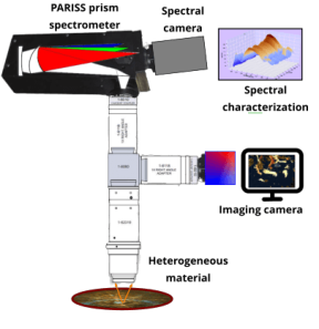
A modular prism based imaging spectrometer and spectrograph captures low signal- to-noise, spatially resolved
spectra, at all wavelengths from 365-nm – 920-nm simultaneously.
When used with a CCD or CMOS camera spectrum detector it becomes an imaging spectrometer. Enables
point-to point spectral imaging in a variety of configurations.
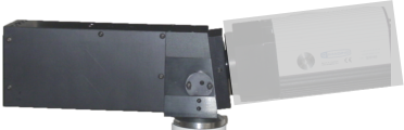
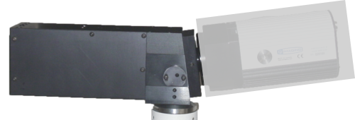
Design: The PARISS imaging spectrograph and spectrometer uses a prism with curved sides to deliver state of
the art light throughput efficiency. This enables highest sensitivity and very fast acquisition times, even with low
signal to noise spectra.
Zoom magnification is available from less than 1x (de-magnification) to 40x, or higher with microscope
objectives.
Most PARISS modules are available separately. Our goal is to enable any researcher to mix-and-match and buy
only what is needed. When budgets are squeezed this is a great way to save money.
Upgrade any time as funds become available.
Mounting: PARISS imaging spectrograph can be column mounted on a bench, a tripod, or interface with a
microscope or telescope video port.
Light collection optics can include a c-mount macro lens, with or without zoom capabilities, a microscope
objective or telescope optics.
Spectral object characterization in %reflection, absorption, or luminescence
Spectral cameras: can be user-supplied or select from a range of options available through LightForm.
Software: Written in Python, various options are available, including:
Basic spectral analysis %Refection, absorption, emission, Spectral classification, Create spectral libraries,
Perform spectral recognition.
Click here for a performance comparison between a prism and a diffraction grating.


PARISS® Modular Imaging Spectrometer And Spectrograph


PARISS® Modular Imaging
Spectrometer And Spectrograph
The PARISS Imaging Spectrometer
is prism-based for the highest
possible spectral sensitivity.
PARISS Imaging
Spectrometer/Spectrograph Overview
A modular prism based imaging spectrometer and
spectrograph captures low signal- to-noise, spatially
resolved spectra, at all wavelengths from 365-nm –
920-nm simultaneously.
When used with a CCD or CMOS camera spectrum
detector it becomes an imaging spectrometer.
Enables point-to point spectral imaging in a variety of
configurations.
Design: The PARISS imaging spectrograph and
spectrometer uses a prism with curved sides to
deliver state of the art light throughput efficiency.
This enables highest sensitivity and very fast
acquisition times, even with low signal to noise
spectra.
Zoom magnification is available from less than 1x
(de-magnification) to 40x, or higher with microscope
objectives.
Most PARISS modules are available separately. Our
goal is to enable any researcher to mix-and-match
and buy only what is needed. When budgets are
squeezed this is a great way to save money.
Upgrade any time as funds become available.
Mounting: PARISS imaging spectrograph can be
column mounted on a bench, a tripod, or interface
with a microscope or telescope video port.
Light collection optics can include a c-mount macro
lens, with or without zoom capabilities, a microscope
objective or telescope optics.
Spectral object characterization in %reflection,
absorption, or luminescence
Spectral cameras: can be user-supplied or select
from a range of options available through LightForm.
Software: Written in Python, various options are
available, including:
Basic spectral analysis %Refection, absorption,
emission, Spectral classification, Create spectral
libraries, Perform spectral recognition.
Zoom magnification: From less than 1x to greater
than 40x. Eliminates the need for multiple
objectives
Samples and specimens: Designed to work with
heterogeneous samples in air, a petri dish,
microscope slide, in water or in-situ
Optics and objectives: Accommodates high
numerical aperture (NA) long working distance
objectives, commercial microscope objectives,
camera, and long focal length telescope lenses for
remote sensing.
Spectroscopy modes: %reflection, %transmission,
absorption fluorescence, luminescence…
Wavelength range: prism-based imaging spectrograph.
Operates from 360 – 920-nm depending on the
spectrum camera and application optics.
Mounts: Can be mounted on a column, microscope C-
mount video port, a cart, tripod, or mast
Illumination: LED brightfield, darkfield, refection,
transmission, monochromatic fluorescence excitation
Observed field-of-view: Coaxial side camera records the
FOV live. Great for object targeting and documentation
PARISS Specifications
Weight: 1,250 g (Excluding a camera) Moving parts: None. Optimizes stability and reproducibility. Dimensions: 210 x 55 x 85 mm Wavelength dispersive element: The wavelength dispersive element is a prism with optical “power.” Concave and convex surfaces on the front and rear surfaces correct astigmatism, coma, and spherical aberration. (See Figure 2) Spectral range: 365 to ~920 nm or 400 to ~920 nm, depending on choice of camera. All spectra acquired simultaneously without order sorting filters Light throughput efficiency: Internal transmission ~90% from 450 to ~920 nm. Entrance slit dimensions: Standard 5 mm by 25- micron, widths of 50 and 100-micron are available in pre-aligned mounting assemblies. Spatial resolution at the sample: Depends on slit width and camera pixel size ~ 0.6 micron by ~0.6 micron with 40x magnification typical. Nanoparticles may be detected but not resolved Spectral resolution: ~1 nm measured at the full width at half maximum of the 436 nm Hg line, depends on slit width and camera pixel size. Optional calibration standards: Available MIDL wavelength calibration lamp and a “SYLPH” NIST certified radiometric light source.