PARISS® Hyperspectral Microscopy
The PARISS Model “PHSI” operates in field
scanning mode in darkfield scatter,
fluorescence, luminescence,
%transmission, %reflection and absorption.
Hyperspectral microscopy work flow
•
Prism based imaging spectrometer for extended wavelength range and light throughput (prism vs
diffraction grating details)
•
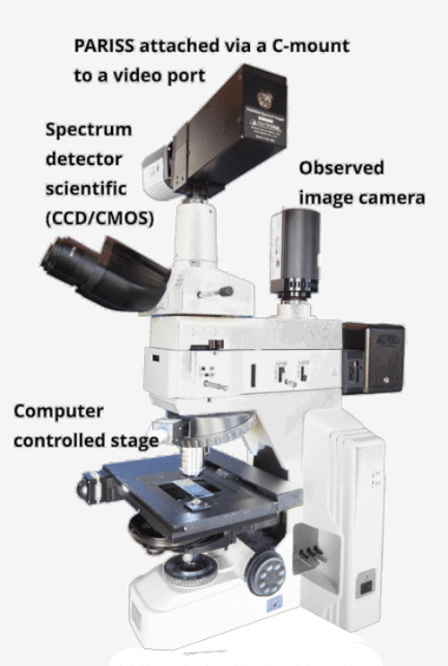
Mounting interface: “C” mount to most research upright and inverted microscopes
•
Wavelength range: 360 to 920-nm simultaneously
•
Spectral resolution: ~1-nm at 436-nm
PARISS imaging spectrometer (Details)
•
Scientific CCD/CMOS spectrum detector
•
Observed image scientific CCD/CMOS
•
Data processor
•
Computer operated microscope stage
•
Custom hyperspectral microscopy software. See here
•
Includes a MIDL calibration lamp to validate wavelength accuracy
•
Optional: NIST certified light source.
•
Optional Xenon illuminator for darkfield nanoparticle characterization
•
Calibration standards: Available MIDL wavelength calibration lamp and a “SYLPH” NIST certified radiometric
light source.
•
Sold either as an accessory to an existing microscope or as a complete system, contact LightForm.
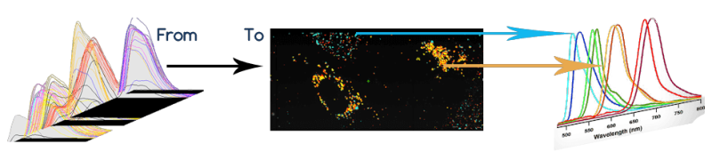
Hyperspectral microscopy correlates spectra presented by the field of view (FOV) with spectra in a reference
spectral library (RSL). An unlimited FOV is acquired by placing a sample on a slide mounted on a computer
controlled translation stage on a microscope. The sample is illuminated with white light for darkfield scatter,
%transmission or %reflection, or with a laser for fluorescence or other excited states. To see a video that
describes how the PARISS hyperspectral microscope works click here.
Each object in the field of view will present a spectrum. If there are tens of thousands of objects, then
PARISS will acquire tens of thousands of spectra many of which are common. The challenge associated with
handling thousands of unique and often common spectra is handled with custom software that sorts and
classifies all spectra in the FOV.
All or some classes of spectra can then be entered into an RSL. Each library spectrum can be pseudo-
colored and linked to a unique target “spectral object”. The presence of one or more pseudo-colors then
confirms the location and presence of a target object.
Future samples can then be scanned, and the spectra presented by all objects in the FOV will be correlated
with those in the library. Those that meet a minimum correlation coefficient will then be “painted” onto a gray
scale image. Various data processing options including counting correlated objects and various mathematical
functions.
Hyperspectral imaging basics
The PARISS hyperspectral microscope acquires many thousands of spectra over an unlimited field of view
(FOV). The PARISS software evaluates all spectra and sorts them into “classes.” One class will likely be
“background spectra,” the remaining spectra will be target objects.
Some, or all, classes of spectra can be added into an RSL. Each library spectrum will be pseudo colored either
by the software or the instrument user.
All, or selected spectra from the FOV that correlate with a library spectrum, will then be “painted” with pixel-
perfect accuracy onto a grayscale image of the FOV.


PARISS® Hyperspectral
Microscopy


The PARISS Model “PHSI” operates in field
scanning mode in darkfield scatter,
fluorescence, luminescence,
%transmission, %reflection and absorption.
•
Scientific CCD/CMOS spectrum detector
•
Observed image scientific CCD/CMOS
•
Data processor
•
Computer operated microscope stage
•
Custom hyperspectral microscopy software.
See here
•
Includes a MIDL calibration lamp to validate
wavelength accuracy
•
Optional: NIST certified light source.
•
Optional Xenon illuminator for darkfield
nanoparticle characterization
•
Calibration standards: Available MIDL
wavelength calibration lamp and a “SYLPH”
NIST certified radiometric light source.
•
Sold either as an accessory to an existing
microscope or as a complete system, contact
LightForm.
Hyperspectral microscopy correlates spectra
presented by the field of view (FOV) with spectra
in a reference spectral library (RSL). An
unlimited FOV is acquired by placing a sample
on a slide mounted on a computer controlled
translation stage on a microscope. The sample is
illuminated with white light for darkfield scatter,
%transmission or %reflection, or with a laser for
fluorescence or other excited states. To see a
video that describes how the PARISS
hyperspectral microscope works click here.
Each object in the field of view will present a
spectrum. If there are tens of thousands of
objects, then PARISS will acquire tens of
thousands of spectra many of which are
common. The challenge associated with handling
thousands of unique and often common spectra
is handled with custom software that sorts and
classifies all spectra in the FOV.
All or some classes of spectra can then be
entered into an RSL. Each library spectrum can
be pseudo-colored and linked to a unique target
“spectral object”. The presence of one or more
pseudo-colors then confirms the location and
presence of a target object.
Future samples can then be scanned, and the
spectra presented by all objects in the FOV will
be correlated with those in the library. Those that
meet a minimum correlation coefficient will then
be “painted” onto a gray scale image. Various
data processing options including counting
correlated objects and various mathematical
functions.
Hyperspectral imaging basics
PARISS imaging spectrometer (Details)
•
Prism based imaging spectrometer for
extended wavelength range and light
throughput (prism vs diffraction grating
details)
•
Mounting interface: “C” mount to most
research upright and inverted microscopes
•
Wavelength range: 360 to 920-nm
simultaneously
•
Spectral resolution: ~1-nm at 436-nm

